Table of Contents
Chapter 3. Elementary Tissues of The Human Body
SHORT ESSAYS (5M)
1.Classify tissues with examples.
Answer:
• Epithelial: Is made of cells arranged in a continuous sheet with one or more layers, has apical & basal surfaces.
o A basement membrane is the attachment between the basal surface of the cell & the underlying connective tissue.
o Two types of epithelial tissues:
(1) Covering & lining epithelia and
(2) Glandular Epithelium.
o The number of cell layers & the shape of the cells in the top layer can classify epithelium.
▪ Simple Epithelium – one cell layer
▪ Stratified epithelium – two or more cell layers
▪ Pseudostratified Columnar Epithelium – When cells of an epithelial tissue are all anchored to the basement Membrane but not all cells reach the apical surface.
▪ Glandular Epithelium – (1) Endocrine: Release hormones directly into the blood stream and
(2) Exocrine – Secrete into ducts.
• Connective: contains many different cell types including fibroblasts, macrophages, mast cells, and adipocytes. Connective Tissue Matrix is made of two materials: ground substance – proteins and polysaccharides, fiber – reticular, collagen, and elastic.
Classification of Connective Tissue:
o Loose Connective – fibers & many cell types in gelatinous matrix, found in skin, & surrounding blood vessels, nerves, and organs.
o Dense Connective – Bundles of parallel collagen fibers& fibroblasts, found in tendons& ligaments. o Cartilage – Cartilage is made of collagen & elastin fibers embedded in a matrix glycoprotein & cells called chondrocytes, which was found in small spaces.
o Cartilage has three subtypes:
▪ Hyaline cartilage – Weakest, most abundant type, Found at end of long bones, & structures like the ear and nose,
▪ Elastic cartilage- maintains shape, branching elastic fibers distinguish it from hyaline and
▪ Fibrous Cartilage – Strongest type, has dense collagen & little matrix, found in pelvis, skull & vertebral discs.
• Muscle: is divided into 3 categories, skeletal, cardiac, and smooth.
o Skeletal Muscle – voluntary, striated, striations perpendicular to the muscle fibers, and it is mainly found attached to bones.
o Cardiac Muscle – involuntary, striated, branched and has intercalated discs o Smooth Muscle – involuntary, nonstriated, spindle-shaped and is found in blood vessels & the GI tract.
• Nervous tissue detects changes in a variety of conditions inside and outside the body and responds by generating action potentials (nerve impulses) that activate muscular contractions and glandular secretions.
2.Explain the differences among skeletal, smooth and cardiac muscles.
Answer:
Skeletal muscle
This type is described as skeletal because it forms those muscles that move the bones (of the skeleton), striated because striations (stripes) can be seen on microscopic examination and voluntary as it is under conscious control. Although most skeletal muscle moves bones, the diaphragm is made from this type of muscle to accommodate a degree of voluntary control in breathing. These fibres (cells) are cylindrical, contain several nuclei and can be up to 35 cm long. Skeletal muscle contraction is stimulated by motor nerve impulses originating in the brain or spinal cord and ending at the neuromuscular junction.
Smooth muscle
Smooth muscle is also described as non-striated, visceral or involuntary. It does not have striations and is not under conscious control. Some smooth muscle has the intrinsic ability to initiate its own contractions (automaticity), e.g. peristalsis. It is innervated by the autonomic nervous system. Additionally, autonomic nerve impulses, some hormones, and local metabolites stimulate its contraction. A degree of muscle tone is always present, meaning that smooth muscle is only completely relaxed for short periods. Contraction of smooth muscle is slower and more sustained than skeletal muscle. It is found in the walls of hollow organs:
• regulating the diameter of blood vessels and parts of the respiratory tract
• propelling contents along, e.g. the ureters, ducts of glands, and the alimentary tract
• expelling contents of the urinary bladder and uterus.
Cardiac muscle
This is only found only in the heart wall. It is not under conscious control but, when viewed under a microscope, cross-stripes (striations) characteristic of skeletal muscle can be seen. Each fiber (cell) has a nucleus and one or more branches. The ends of the cells and their branches are in very close contact with the ends and branches of adjacent cells. Microscopically these ‘joints’, or intercalated discs, appear as lines that are thicker and darker than the ordinary cross-stripes. This arrangement gives cardiac muscle the appearance of a sheet of muscle rather than a very large number of individual fibers.
3.Define cartilage, mention its type. Give the description, function, and examples of each of them.
Answer: Cartilage is firmer than other connective tissues. The cells (chondrocytes) are sparse and lie embedded in a matrix reinforced by collagen and elastic fibers. There are three types: hyaline cartilage, fibrocartilage, and elastic fibrocartilage.
Hyaline cartilage
Hyaline cartilage is a smooth bluish-white tissue. The chondrocytes are arranged in small groups within cell nests and the matrix is solid and smooth. Hyaline cartilage provides flexibility, support, and smooth surfaces for movement at joints. It is found:
• on the ends of long bones that form joints
• forming the costal cartilages, which attach the ribs to the sternum
• forming part of the larynx, trachea, and bronchi.
Fibrocartilage
This consists of dense masses of white collagen fibers in a matrix similar to that of hyaline cartilage with the cells widely dispersed. It is a tough, slightly flexible, supporting tissue found:
• as pads between the bodies of the vertebrae, the intervertebral discs
• between the articulating surfaces of the bones of the knee joint, called semilunar cartilages
• on the rim of the bony sockets of the hip and shoulder joints, deepening the cavities without restricting movement.
Elastic fibrocartilage
This flexible tissue consists of yellow elastic fibers lying in a solid matrix with chondrocytes lying between the fibers. It provides support and maintains the shape of, e.g. the pinna or lobe of the ear, the epiglottis, and part of the tunica media of blood vessel walls.
4.Differentiate between loose and dense connective tissues.
Answer:
Dense connective tissue
This contains more fibers and fewer cells than loose connective tissue.
➢ Fibrous tissue
This tissue is made up mainly of closely packed bundles of collagen fibers with very little matrix. Fibrocytes
(old and inactive fibroblasts) are few in number and lie in rows between the bundles of fibers. Fibrous tissue
is found:
• forming ligaments, which bind bones together
• as an outer protective covering for bone, called the periosteum
• as an outer protective covering of some organs, e.g. the kidneys, lymph nodes, and the brain
• forming muscle sheaths, called muscle fascia, which extend beyond the muscle to become the tendon that attaches the muscle to bone.
➢ Elastic tissue
Elastic tissue is capable of considerable extension and recoil. There are few cells and the matrix consists mainly of masses of elastic fibers secreted by fibroblasts. It is found in organs where stretching or alteration of shape is required, e.g. in large blood vessel walls, the trachea and bronchi, and the lungs.
Loose connective tissue
This is the most generalized type of connective tissue. The matrix is semisolid with many fibroblasts and some fat cells (adipocytes), mast cells, and macrophages widely separated by elastic and collagen fibers. It is found in almost every part of the body, providing elasticity and tensile strength. It connects and supports other tissues, for example:
• under the skin
• between muscles
• supporting blood vessels and nerves
• in the alimentary canal
• in glands supporting secretory cells.
5.Describe the different types of epithelial tissues with examples.
Answer: Simple epithelia & Stratified epithelia
Squamous (pavement) epithelium
This is composed of a single layer of flattened cells. The cells fit closely together like flat stones, forming a thin and very smooth membrane across which diffusion occurs easily. It forms the lining of the following structures:
➢ heart – where it is known as the endocardium
➢ blood vessels where it is also known
➢ lymph vessels as the endothelium
➢ alveoli of the lungs
➢ lining the collecting ducts of nephrons in the kidneys
Cuboidal epithelium
This consists of cube-shaped cells fitting closely together lying on a basement membrane. It forms the kidney tubules and is found in some glands such as the thyroid. Cuboidal epithelium is actively involved in secretion, absorption, and/or excretion.
Columnar epithelium
This is formed by a single layer of cells, rectangular in shape, on a basement membrane. It lines many organs and often has adaptations that make it well suited to a specific function. The lining of the stomach is formed from simple columnar epithelium without surface structures. The free surface of the columnar epithelium lining the small intestine is covered with microvilli.
Stratified epithelia
Stratified epithelia consist of several layers of cells of various shapes. Continual cell division in the lower (basal) layers pushes cells above nearer and nearer to the surface, where they are shed. Basement membranes are usually absent. The main function of stratified epithelium is to protect underlying structures from mechanical wear and tear. There are two main types: stratified squamous and transitional.
Stratified squamous epithelium
This is composed of several layers of cells. In the deepest layers, the cells are mainly columnar and, as they grow towards the surface, they become flattened and are then shed.
Keratinized stratified epithelium. This is found on dry surfaces subjected to wear and tear, i.e. skin, hair, and nails. The surface layer consists of dead epithelial cells that have lost their nuclei and contain the protein keratin. This forms a tough, relatively waterproof protective layer that prevents drying of the live cells underneath. The surface layer of skin is rubbed off and is replaced from below.
Non-keratinized stratified epithelium. This protects moist surfaces subjected to wear and tear and prevents them from drying out, e.g. the conjunctiva of the eyes, the lining of the mouth, the pharynx, the esophagus, and the vagina.
Transitional epithelium
This is composed of several layers of pear-shaped cells. It lines several parts of the urinary tract including the bladder and allows for stretching as the bladder fills.
6.Explain the structure and functions of neurons.
Answer:
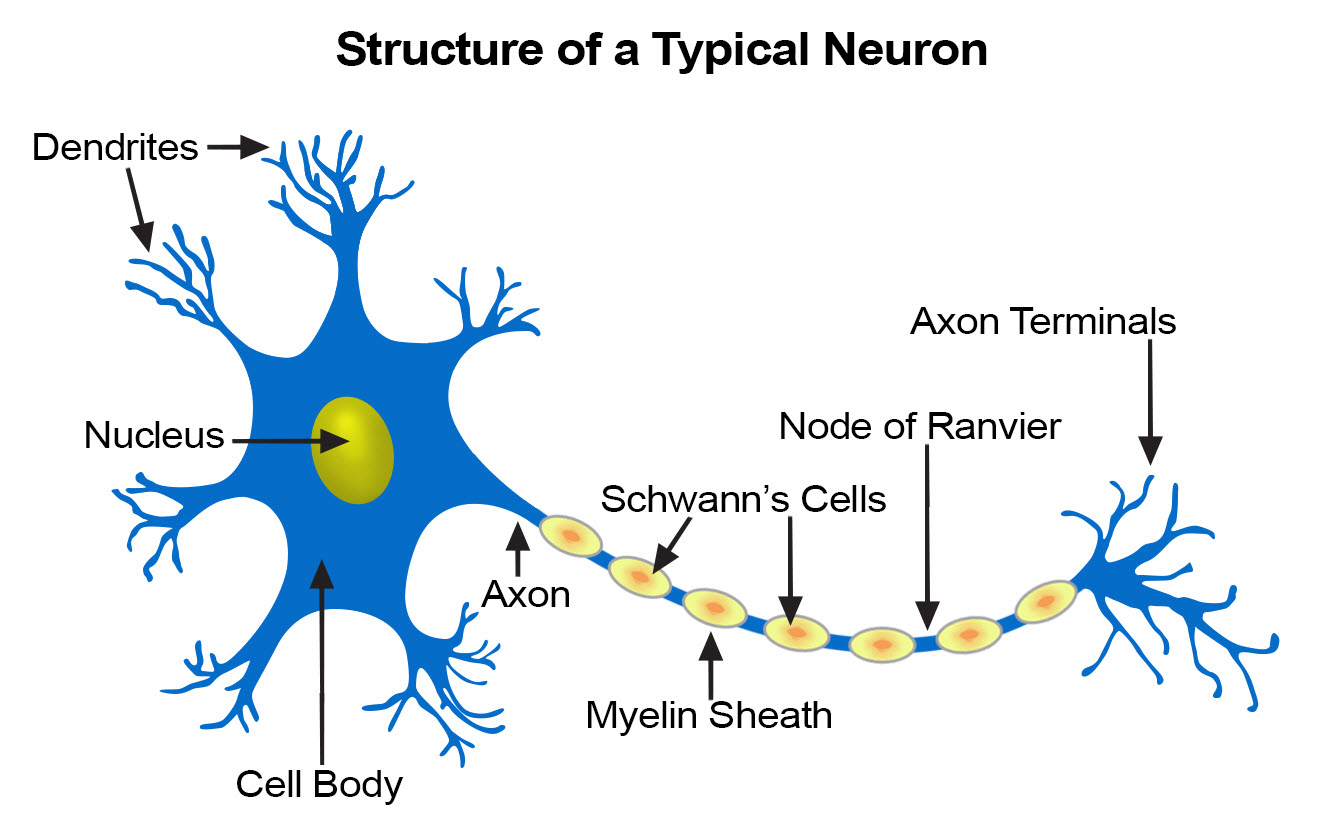
Each neuron consists of a cell body and its processes, one axon, and many dendrites. Neurons are commonly referred to as nerve cells. Bundles of axons bound together are called nerves. Neurons cannot divide, and for survival, they need a continuous supply of oxygen and glucose. Unlike many other cells, neurons can synthesize chemical energy (ATP) only from glucose.
Neurons generate and transmit electrical impulses called action potentials. The initial strength of the impulse is maintained throughout the length of the neuron. Some neurons initiate nerve impulses while others act as ‘relay stations’ where impulses are passed on and sometimes redirected. Nerve impulses can be initiated in response to stimuli from:
• outside the body, e.g. touch, light waves
• inside the body, e.g. a change in the concentration of carbon dioxide in the blood alters respiration; a thought may result in involuntary movement.
SHORT ANSWERS (2M)
1. Classify muscular tissue with examples.
Answer: Muscle: is divided into 3 categories, skeletal, cardiac, and smooth.
o Skeletal Muscle – voluntary, striated, striations perpendicular to the muscle fibers, and it is mainly found attached to bones.
o Cardiac Muscle – involuntary, striated, branched, and has intercalated discs
o Smooth Muscle – involuntary, nonstriated, spindle-shaped and is found in blood vessels & the GI tract.
2. Classify connective tissue with examples.
Answer: Connective: contains many different cell types including fibroblasts, macrophages, mast cells, and adipocytes. Connective Tissue Matrix is made of two materials: ground substance – proteins and polysaccharides, fiber – reticular, collagen and elastic.
Classification of Connective Tissue:
o Loose Connective – fibers & many cell types in a gelatinous matrix, found in skin, & surrounding blood vessels, nerves, and organs.
o Dense Connective – Bundles of parallel collagen fibers& fibroblasts, found in tendons& ligaments.
o Cartilage – Cartilage is made of collagen & elastin fibers embedded in a matrix glycoprotein & cells called chondrocytes, which were found in small spaces.
o Cartilage has three subtypes:
▪ Hyaline cartilage – Weakest, most abundant type, Found at end of long bones, & structures like the ear and nose,
▪ Elastic cartilage– maintains shape, branching elastic fibers distinguish it from hyaline and
▪ Fibrous Cartilage – Strongest type, has dense collagen & little matrix, found in pelvis, skull & vertebral discs
3. Draw a neat labeled diagram of neurons.
4. Classify simple epithelium with examples.
Answer: Squamous (pavement) epithelium or Simple epithelium
This is composed of a single layer of flattened cells. The cells fit closely together like flat stones, forming a thin and very smooth membrane across which diffusion occurs easily. It forms the lining of the following structures:
➢ heart – where it is known as the endocardium
➢ blood vessels where it is also known
➢ lymph vessels as the endothelium
➢ alveoli of the lungs
➢ lining the collecting ducts of nephrons in the kidneys
Cuboidal epithelium
This consists of cube-shaped cells fitting closely together lying on a basement membrane. It forms the kidney tubules and is found in some glands such as the thyroid. Cuboidal epithelium is actively involved in secretion, absorption and/or excretion.
Columnar epithelium
This is formed by a single layer of cells, rectangular in shape, on a basement membrane. It lines many organs and often has adaptations that make it well suited to a specific function. The lining of the stomach is formed from simple columnar epithelium without surface structures. The free surface of the columnar epithelium lining the small intestine is covered with microvilli.
5. Classify compound epithelium with examples.
Answer: Stratified epithelia or Compound epithelium
Stratified epithelia consist of several layers of cells of various shapes. Continual cell division in the lower (basal) layers pushes cells above nearer and nearer to the surface, where they are shed. Basement membranes are usually absent. The main function of stratified epithelium is to protect underlying structures from mechanical wear and tear.
There are two main types: stratified squamous and transitional.
Stratified squamous epithelium
This is composed of several layers of cells. In the deepest layers, the cells are mainly columnar and, as they grow towards the surface, they become flattened and are then shed.
Keratinized stratified epithelium. This is found on dry surfaces subjected to wear and tear, i.e. skin, hair and nails. The surface layer consists of dead epithelial cells that have lost their nuclei and contain the protein keratin. This form a tough, relatively waterproof protective layer that prevents drying of the live cells underneath. The surface layer of skin is rubbed off and is replaced from below.
Non-keratinized stratified epithelium. This protects moist surfaces subjected to wear and tear, and prevents them from drying out, e.g. the conjunctiva of the eyes, the lining of the mouth, the pharynx, the oesophagus and the vagina.
Transitional epithelium
This is composed of several layers of pear-shaped cells. It lines several parts of the urinary tract including the bladder and allows for stretching as the bladder fills.
6. Write the general characteristics of epithelial tissue.
Answer: This tissue type covers the body and lines cavities, hollow organs, and tubes. It is also found in glands. The structure of epithelium is closely related to its functions, which include:
• protection of underlying structures from, for example, dehydration, chemical, and mechanical damage
• secretion
• absorption.
The cells are very closely packed and the intercellular substance, the matrix, is minimal. The cells usually lie on a basement membrane, which is an inert connective tissue made by the epithelial cells themselves. Epithelial tissue may be:
• simple: a single layer of cells
• stratified: several layers of cells.
7. Write the location and functions of the Transitional epithelium.
Answer: Transitional epithelium
This is composed of several layers of pear-shaped cells. It lines several parts of the urinary tract including the bladder and allows for stretching as the bladder fills.
Also Read :
Chapter 1. Scope of anatomy and physiology, basic terminologies used in this subject
Chapter 2. Structure of cell – its components and their functions.


