
Table of Contents
SHORT ESSAYS (5M)
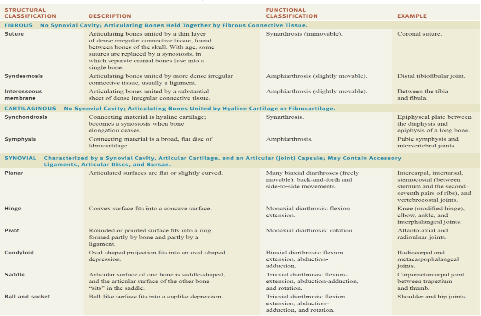
1. Classify joints with examples
Answer:
2. Explain the Haversian canal system of bone.
Answer: Haversian canals a series of microscopic tubes in the outermost region of bone called cortical bone that allows blood vessels and nerves to travel through them. Each Haversian canal generally contains one or two capillaries and nerve fibers. The channels are formed by concentric layers called lamellae. The Haversian canals surround blood vessels and nerve cells throughout bones and communicate with bone cells (contained in spaces within the dense bone matrix called lacunae) through connections called canaliculi. This unique arrangement is conducive to mineral salt deposits and storage which gives bone tissue its strength.
In mature compact bone, most of the individual lamellae form concentric rings around larger longitudinal canals within the bone tissue. These canals are called Haversian canals. Haversian canals are contained within osteons, which are typically arranged along the long axis of the bone in parallel to the surface. The canals and the surrounding lamellae (8-15) form the functional unit, called a Haversian system or osteon.
3. Write the composition and functions of bone.
Answer: Function of bone
I. Providing the body framework
II. Giving attachment to muscles and tendons
III. Allowing movement of the body as a whole and of parts of the body, by forming joints that are moved by muscles
IV. Forming the boundaries of the cranial, thoracic, and pelvic cavities, and protecting the organs they contain
V. Haemopoiesis, the production of blood cells in red bone marrow
VI. Mineral storage, especially calcium phosphate the mineral reservoir within the bone is essential for the maintenance of blood calcium levels, which must be tightly controlled.
Composition of bone
Osteoblasts
These bone-forming cells are responsible for the deposition of both inorganic salts and osteoid in bone tissue.
They are therefore present at sites where the bone is growing, repairing or remodelling, e.g.:
a) In the deeper layers of the periosteum
b) In the centres of ossification of immature bone
c) At the ends of the diaphysis adjacent to the epiphyseal cartilages of long bones
d) At the site of a fracture.
As they deposit new bone tissue around themselves, they eventually become trapped in tiny pockets (lacunae) in the growing bone and differentiate into osteocytes.
Osteocytes
These are mature bone cells that monitor and maintain bone tissue and are nourished by tissue fluid in the canaliculi that radiate from the central canals.
Osteoclasts
These cells break down bone, releasing calcium and phosphate. They are very large cells with up to 50 nuclei, which have formed from the fusion of many monocytes. The continuous remodeling of healthy bone tissue is the result of a balanced activity of the bone’s osteoblast and osteoclast populations. Osteoclasts are found in areas of the bone where there is active growth, repair, or remodeling,
e.g.:
a) Under the periosteum, maintaining bone shape during growth and removing excess callus formed during the 0healing of fractures.
b) Round the walls of the medullary canal during growth and to canalize callus during healing.
4. Describe the angular and special movements of joints.
Answer: Special movements
a) Elevation is an upward movement of a part of the body, such as closing the mouth at the temporomandibular joint (between the mandible and temporal bone) to elevate the mandible.
b) Depression is a downward movement of a part of the body, such as opening the mouth to depress the mandible or returning shrugged shoulders to the anatomical position to depress the scapula.
c) Protraction is a movement of a part of the body anteriorly in the transverse plane.
d) Retraction is a movement of a protracted part of the body back to the anatomical position.
e) Inversion is the movement of the sole medially at the intertarsal joints (between the tarsal).
f) Eversion is a movement of the sole laterally at the intertarsal joints.
g) Dorsiflexion refers to bending of the foot at the ankle or talocrural joint in the direction of the dorsum. Dorsiflexion occurs when you stand on your heels.
h) Plantar flexion involves bending of the foot at the ankle joint in the direction of the plantar or inferior surface, as when you elevate your body by standing on your toes.
i) Supination is a movement of the forearm at the proximal and distal radioulnar joints in which the palm is turned anteriorly.
j) Pronation is a movement of the forearm at the proximal and distal radioulnar joints in which the distal end of the radius crosses over the distal end of the ulna and the palm is turned posteriorly.
k) Opposition is the movement of the thumb at the carpometacarpal joint in which the thumb moves across the palm to touch the tips of the fingers on the same hand
Angular movements (Increase or decrease in the angle between bones)
a) Flexion – Decrease in the angle between articulating bones, usually in the sagittal plane.
b) Lateral flexion – Movement of the trunk in the frontal plane.
c) Extension – Increase in the angle between articulating bones, usually in the sagittal plane.
d) Hyperextension – Extension beyond the anatomical position.
e) Abduction – Movement of a bone away from the midline, usually in the frontal plane.
f) Adduction – Movement of a bone toward the midline, usually in the frontal plane.
g) Circumduction – Flexion, abduction, extension, and adduction in succession, in which the distal end of a body part moves in a circle.
5. What is a synovial joint? Explain its types with examples.
Answer: Synovial joints are characterized by the presence of a space or capsule between the articulating bones. The ends of the bones are held close together by a sleeve of fibrous tissue and lubricated with a small amount of fluid. Synovial joints are the most moveable of the body.
|
Synovial Joint Types (structural classification) |
Description |
Example |
|
Planar |
Articulated surfaces are flat or slightly curved | Intercarpal, Intertarsal, Sternocostal (between the sternum and the second– seventh pairs of ribs), and vertebrocostal joints |
|
Hinge |
Convex surface fits into a concave surface | Knee (modified hinge), extension. elbow, ankle, and interphalangeal joints |
|
Pivot |
A rounded or pointed surface fits into a ring.
formed partly by bone and partly by a ligament. |
Atlanto-axial and radioulnar joints. |
|
Condyloid |
Oval-shaped projection fits into an oval-shaped depression. | Radiocarpal and metacarpophalangeal joints. |
|
Saddle |
The articular surface of one bone is saddle-shaped, and the articular surface of the other bone “sits” in the saddle. | The carpometacarpal joint between trapezium and thumb. |
|
Ball-and-socket |
A ball-like surface fits into a cuplike depression. | Shoulder and hip joints. |
6.Classify the axial skeletal system with examples.
Answer: In the human skeleton, axial skeletal consists of 80 bones and is composed of six parts; the skull bones, the ossicles of the middle ear, the hyoid bone, the rib cage, sternum and the vertebral column.
|
Facial Bones |
Auditory Ossicles |
|
|
|||||
|
|
|
|
|
|||||
|
Temporal (2) |
Zygomatic (2) |
Incus (2) |
Thoracic vertebrae (12) |
Ribs (24) |
|||||
|
|
|
|
||||||
|
|
Sacrum (1) |
|||||||
|
|
|
|||||||
|
|
||||||||
|
|||||||||
|
7.Classify the appendicular skeletal system with examples.
Answer:
|
Pectoral girdles |
Upper Extremity |
Pelvic Girdle |
Lower Extremity |
|
|
Clavicle (2) |
Humerus (2) |
Coxal, innominate, or hip bones (2) |
Femur (2) |
|
|
Scapula (2) |
Radius (2) |
Tibia (2) | ||
|
Ulna (2) |
Fibula (2) |
|||
| Carpals (16) | Patella (2) | |||
| Metacarpals (10) | Tarsals (14) | |||
| Phalanges (28) | Metatarsals (10) | |||
| Phalanges (28) | ||||
8.Classify bones on the basis of shape along with examples.
Answer:
| 1. | Long Bones | “Long bones” are longer than they are wide, i.e. length > diameter. Long bones are usually somewhat curved – contributing to their mechanical strength. |
• • Femur (leg bone) • • Tibia (leg bone) • • Fibula (leg bone) • • Humerus (arm bone) • • Ulna (arm bone) • • Radius (arm bone)
|
| 2. | Short Bones | “Short bones” can be approximately cube-shaped, i.e. length is similar to width/depth/diameter. | • Scaphoid bone (wrist bone)
• Lunate bone (wrist bone) • Hamate bone (wrist bone) and other wrist bones = carpal bones • Cuboid bone (ankle bone) • First Cuniform bone (ankle bone) • Second Cuniform bone (ankle bone) and other ankle bones = tarsal bones
|
| 3. | Flat Bones | “Flat bones” have a thin shape and, in some cases, provide mechanical protection to soft tissues beneath or enclosed by the flat bone e.g. cranial bones that protect the brain. Flat bones also have extensive surfaces for muscle attachments e.g. scapulae (shoulder) bones. |
• Cranial bones (protecting the brain) e.g. o Parietal bones
• Sternum (protecting organs in the thorax) • Ribs (protecting organs in the thorax) • Scapulae (shoulder blades).
|
| 4. | Irregular Bones | “Irregular bones” have complicated shapes that cannot be classified as “long”, “short” or “flat”. Their shapes are due to the functions they fulfill within the body e.g. providing major mechanical support for the body yet also protecting the spinal cord (in the case of the vertebrae). |
• Atlas bone • Axis bone and other vertebrae • Hyoid bone • Sphenoid bone • Zygomatic bones and other facial bones. |
| 5. | Sesamoid Bones | “Sesamoid bones” develop in some tendons in locations where there is considerable friction, tension, and physical stress. | Only one type of sesamoid bone is present in all normal human skeletons so has a name. That is the patella (singular), patellae (plural). Patellae are also called “kneecaps”. Complete human skeletons include 2 of these, one in each leg. |
SHORT ANSWERS (2M)
1. Name synovial joints
Answer: Planar, Hinge, Pivot, Condyloid, Saddle, and Ball-and-socket
2. Classify bone marrow and functions.
Answer:
Bone marrow can be 1 of 2 types, red or yellow, depending on whether it consists of mainly hematopoietic (red-colored) tissue or fatty (yellow-colored) tissue. Bone marrow functions primarily to produce blood cells and to store fat.
Red marrow is found mainly in the flat bones, such as the hip bone, sternum (breast) bone, skull, ribs, vertebrae, and shoulder blades, as well as in the metaphyseal and epiphyseal ends of the long bones, such as the femur, tibia, and humerus, where the bone is cancellous or spongy.
Yellow marrow is found in the hollow interior of the diaphyseal portion or the shaft of long bones. By the time a person reaches old age, nearly all of the red marrow is replaced by yellow marrow. However, the yellow marrow can revert to red if there is increased demand for red blood cells, such as in instances of blood loss.
3. Define an articulation.
Answer: Articulation is the connection made between bones in the body which link the skeletal system into a functional whole.
4. Write the histology of bone
Answer: Bone is a strong and durable type of connective tissue. Its major constituent (65%) is a mixture of calcium salts, mainly calcium phosphate. This inorganic matrix gives bone great hardness, but on its own would be brittle and prone to shattering. The remaining third is an organic material, called osteoid, which is composed mainly of collagen. Collagen is very strong and gives bone slight flexibility. The cellular component of bone contributes less than 2% of bone mass.
Bone cells
There are three types of bone cell:
• Osteoblast
• Osteocyte
• Osteoclast
5. Name the bones of the cranium
Answer: 1 frontal bone, 2 parietal bones, 2 temporal bones, 1 occipital bone, 1 sphenoid bone & 1 ethmoid bone
6. Name the bone cells
Answer: Bone cells
There are three types of bone cell:
• Osteoblast: These bone-forming cells are responsible for the deposition of both inorganic salts and osteoid in bone tissue. They are therefore present at sites where the bone is growing, repairing, or remodeling.
• Osteocyte: These are mature bone cells that monitor and maintain bone tissue, and are nourished by tissue fluid in the canaliculi that radiate from the central canals.
• Osteoclast: These cells break down bone, releasing calcium and phosphate. They are very large cells with up to 50 nuclei, which have formed from the fusion of many monocytes
7. Name the bones of the face.
Answer: 2 zygomatic (cheek) bones, 1 maxilla, 2 nasal bones, 2 lacrimal bones, 1 vomer, 2 palatine bones, 2 inferior conchae & 1 mandible.
8. Mention the bones of the upper limb.
Answer: Humerus, Ulna, Radius, Carpal (wrist) bones (There are eight carpal bones arranged in two rows of four. From outside inwards they are proximal row: scaphoid, lunate, triquetrum, pisiform and distal row: trapezium, trapezoid, capitate, hamate), Metacarpal bones (bones of the hand) and Phalanges (finger bones).
9. Mention the bones of the lower limb.
Answer: Femur (thigh bone), Tibia (shin bone), Fibula, Patella (knee cap), Tarsal (ankle) bones, Metatarsals (bones of the foot), and Phalanges (toe bones).
10. Name the bones of the vertebral column.
Answer: Cervical vertebrae (7), Thoracic vertebrae (12), Lumbar vertebrae (5), Sacrum (1), and Coccyx (1).
11.Write the functions of bone.
Answer: Function of bone
I. Providing the body framework
II. Giving attachment to muscles and tendons
III. Allowing movement of the body as a whole and of parts of the body, by forming joints that are moved by muscles
IV. Forming the boundaries of the cranial, thoracic, and pelvic cavities, and protecting the organs they contain
V. Haemopoiesis, the production of blood cells in red bone marrow
VI. Mineral storage, especially calcium phosphate the mineral reservoir within the bone is essential for the maintenance of blood calcium levels, which must be tightly controlled.
Also Read:
Chapter 1. Scope of anatomy and physiology, basic terminologies used in this subject
Chapter 2. Structure of cell – its components and their functions.
Chapter 3. Elementary Tissues of The Human Body


[…] Also Read: Chapter – 4. Osseous System – RecNotes […]