
Table of Contents
Simple Staining: Principle, Procedure, Uses
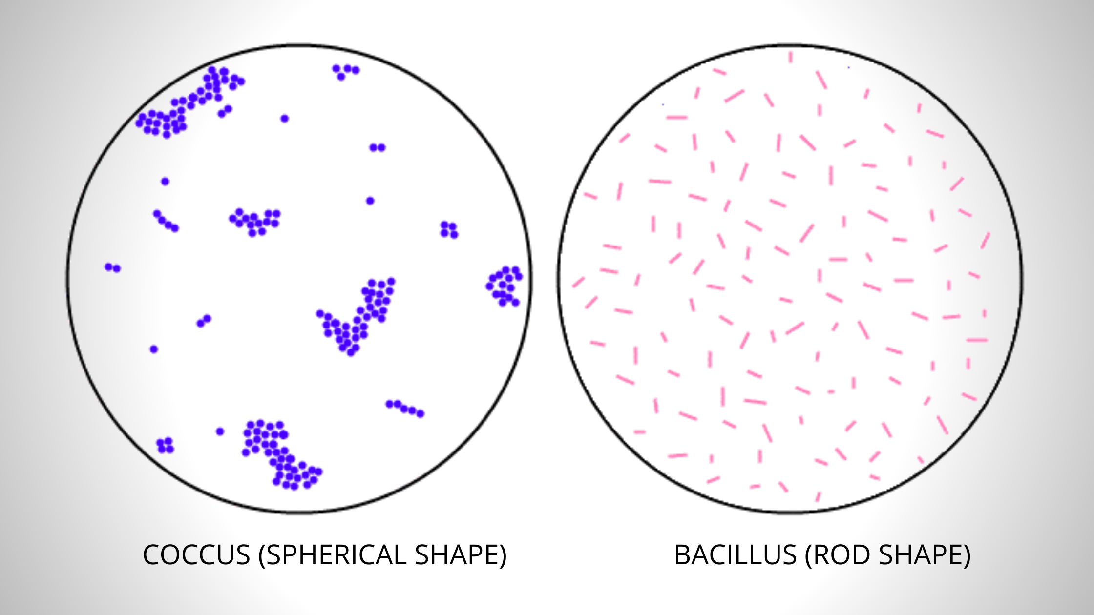
The simple stain can be used as a quick and easy way to determine cell shape, size and arrangements of bacteria. True to its name, the simple stain is a very simple staining procedure involving a single solution of stain. Any basic dye such as methylene blue, safranin, or crystal violet can be used to color the bacterial cells.
These stains will readily give up a hydroxide ion or accept a hydrogen ion, which leaves the stain positively charged. Since the surface of most bacterial cells and cytoplasm is negatively charged, these positively charged stains adhere readily to the cell surface. After staining, bacterial cell morphology (shape and arrangements) can be appreciated.
Procedure

Preparation of a smear

- Using a sterilized inoculating loop, transfer loopful of liquid suspension containing bacteria to a slide (clean grease free microscopic slide) or transfer an isolated colony from a culture plate to a slide with a water drop.
- Disperse the bacteria on the loop in the drop of water on the slide and spread the drop over an area the size of a dime. It should be a thin, even smear.
- Allow the smear to dry thoroughly.

- Heat-fix the smear cautiously by passing the underside of the slide through the burner flame two or three times. It fixes the cell in the slide. Do not overheat the slide as it will distort the bacterial cells.
Staining
- Cover the smear with methylene blue and allow the dye to remain in the smear for approximately one minute (Staining time is not critical here; somewhere between 30 seconds to 2 minutes should give you an acceptable stain, the longer you leave the dye in it, the darker will be the stain).
- Using distilled water wash bottle, gently wash off the excess methylene blue from the slide by directing a gentle stream of water over the surface of the slide.
- Wash off any stain that got on the bottom of the slide as well.
- Saturate the smear again but this time with Iodine. Iodine will set the stain
- Wash of any excess iodine with gently running tap water. Rinse thoroughly. (You may not get a mention of step 4 and 5 in some textbooks)
- Wipe the back of the slide and blot the stained surface with bibulous paper or with a paper towel.
- Place the stained smear on the microscope stage smear side up and focus the smear using the 10X objective.
- Choose an area of the smear in which the cells are well spread in a monolayer. Center the area to be studied, apply immersion oil directly to the smear, and focus the smear under oil with the 100X objective.
Results
The bacterial cells usually stain uniformly and the color of the cell depends on the type of dye used. If methyene blue is used, some granules in the interior of the cells of some bacteria may appear more deeply stained than the rest of the cell, which is due to presence of different chemical substances.
Advantages
- Simple staining is a very simple method to perform, which stains the organism by using a single reagent.
- It is a rapid method that reduces the performance time by taking only 3-5 minutes.
- Simple staining helps to examine or elucidate the bacterial shape, size and arrangement.
- It also helps us to differentiate the bacterial cells from the non-living structures.
- Simple staining can be useful in the preliminary study of the bacteria’s morphological characteristics.
Disadvantages
- It does not give much information about the cell apart from the bacteria’s morphological characteristics.
- Through simple staining, we cannot classify a particular type of organism.
Uses
Diagnostic microbiology laboratory generally does not perform simple staining methods. Differential staining such as Gram Staining and AFB Staining are commonly used to identify and differentiate the bacterial isolates. Simple staining can be useful in the following circumstances.
- To differentiate bacteria from yeast cells: When endocervical swab culture is done in blood agar both Staphylococcus spp and yeast cells may give similar-looking colonies in Blood agar (a common error for a new technologist or microbiologist with less experience). Performing the wet mount technique or simple staining from the isolate can be helpful.
- To presumptively identify the bacterial isolate
Due to their ubiquitous presence, Bacillus spp may present as a contaminant in the culture plates. In some circumstances (e.g. growth in Blood Agar but no growth in MacConkey Agar), identifying the shape of the bacteria (rod or cocci) may help to eliminate the isolate as possible contaminants (e.g., Bacillus spp) or further process as a potential pathogen (cocci).
Conclusion
Therefore, we can conclude that a simple staining method is the easiest way to colour the microscopic object as it uses a single basic stain. The results of simple staining are based on the type of basic stain that has been used.
The colour of a stain will decide the colour of a specimen that has to be identified. For example, when the bacteria retain the safranin colour, they appear pink-red, and the same goes with the other stains.
There is an attraction between the positive stain and the negative bacterial cell in simple staining, which results in the observation of coloured bacteria with a bright background.
Read This Also: Microbiology Exam Guide